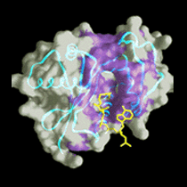
Visualization of Virus Particles
 The Wistar Institute has a major interest in studying the three-dimensional
structure of viruses to understand their properties and evolutionary
relationships. Wistar is collaborating with the University of Helsinki to
use electron microscopy and crystallography to image viruses and their
component molecules. Collaboration between the institutions is done over
the Internet. The most demanding task is exchanging images of complete
virus particles--both the raw 2D EM data and the 3D reconstructions made
from them.
The Wistar Institute has a major interest in studying the three-dimensional
structure of viruses to understand their properties and evolutionary
relationships. Wistar is collaborating with the University of Helsinki to
use electron microscopy and crystallography to image viruses and their
component molecules. Collaboration between the institutions is done over
the Internet. The most demanding task is exchanging images of complete
virus particles--both the raw 2D EM data and the 3D reconstructions made
from them.
although adenoviruses cause human ailments such as respiratory infections,
conjunctivitis and enteric dysentery, they can also be used as vectors in
human gene therapy to combat sickness. The organization of the virion is
being studied using electron microscopy and image analysis. A
three-dimensional image reconstruction at high resolution from
cryo-electron micrographs has revealed how hexon proteins form the viral
facets and showed the interaction of penton base and fiber at the vertex.
Wistar is also attempting to crystallize the entire adenovirus.
although Bacteriophage PRD1 is structurally unusual, recent work has shown
its remarkable similarities to adenovirus. Both are icosahedral with vertex
fibers, have trimeric major coat proteins, and contain double-stranded
linear DNA with terminal proteins, PRD1 is the only known spherical
prokaryotic virus to have this form of genome, and is also unique in
possessing a lipid membrane within its outer capsid. The viral capsid, or
outer shell, is formed from two proteins; P31 lies at the vertices, while
the major coat protein, P3, forms the facets. Wistar is investigating the
PRD1 structure in collaboration with the University of Helsinki, Finland.
The strong similarities between P3 and hexon suggest strongly that PRD1 and
adenovirus are related, and establish the first direct structural link
between viruses from the animal and bacterial kingdoms. The analogy is
suggesting interesting directions for future research, such as the role
that the PRD1 fiber proteins may play in entry. A more detailed portrait of
the PRD1 virion will be obtained by combining the EM and X-ray images. This
will lead to an increased understanding of PRD1 organization and stability,
and allow further exploration of its intriguing similarity to adenovirus.
Collaborators
The Wistar Institute, University of Pennsylvania, USA;
University of Helsinki, Finland
Contact
Roger M. Burnett
The Wistar Institute, University of Pennsylvania, USA
burnett@wistar.upenn.edu
Dennis H. Bamford
Department of Biosciences, University of Helsinki, Finland
dennis.bamford@helsinki.fi
http://www.wistar.upenn.edu