

Real-Time Multi-Scale Brain Data Acquisition, Assembly, and Analysis using an End-to-End OptIPuter
|
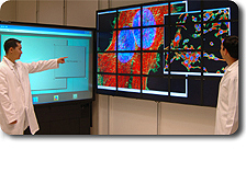
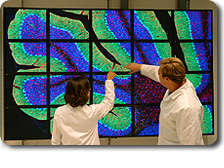
URL: http://ncmir.ucsd.edu/iGrid2005 Contact: David Lee, National Center for Microscopy Imaging Research (NCMIR), University of California, San Diego (UCSD), USA, dlee @ ncmir.ucsd.edu Collaborators: NCMIR, UCSD, USA: David Lee, Mark Ellisman, Ruth West, Maryann Martone, Steve Lamont, Steve Peltier, Tom Hutton, Tom Deerink, Masako Terada, Tomas Molina Electronic Visualization Laboratory, University of Illinois at Chicago (UIC), USA: Jason Leigh, Nicholas Schwarz, Luc Renambot, Andrew Johnson, Byungil (Brent) Jeong, Rajvikram Singh, Venkatram Vishwanath, Eric He, Chaoyue (Joy) Xiong Concurrent Systems Architecture Group, UCSD, USA: Nut Taesombut, Xinran (Ryan) Wu, Eric Weigle, Andrew Chien International Center for Advanced Internet Research, Northwestern University, USA: Joe Mambretti, Jim Chen, Fei Yeh Laboratory for Advanced Computing, UIC, USA: Bob Grossman Cybermedia Center, Osaka University, Japan: Toyokazu Akiyama, Shinji Shimojo, Susumu Research Center for Ultra High Voltage Electron Microscope, Osaka University, Japan: Hirotaro Mori KDDI R&D Laboratories, Japan: Atushi Koike, Hiromu Otsuka, Sei Naito National Center for High Performance Computing, Taiwan: Fang-Pang Lin, Sun-In Lin, Shi-Wei Lo Advanced Internet Research Group, University of Amsterdam, NL: Cees de Laat Korea Institute of Science and Technology Information, Korea: Ge-Bum Koo The operation of a scientific experiment on a global testbed consisting of visualization, storage, computational, and network resources is demonstrated. These resources are bundled into a unified platform using OptIPuter-developed technologies, including dynamic lambda allocation, advanced transport protocols, the Distributed Virtual Computer (DVC) middleware, and a multi-resolution visualization system running over the Scalable Adaptive Graphics Environment (SAGE). With these layered technologies, runs a multi-scale correlated microscopy experiment where a biologist images a sample and progressively magnifies it, zooming from an entire system, such as a rat cerebellum, to an individual spiny dendrite. Using both the 100Megapixel LambdaVision display and the auto-stereo Personal Varrier, scientists can effectively and simultaneously view every step of a multi-scale microscopy correlation process, viewing large 2D scenes and 3D subsections of the scene while comparing them to dozens of possible contexts and matching these to live video output of an electron microscope. |
|